 Ελληνικά
Kidney stone
Kidney stones are one of the most painful urological diseases that have affected people for centuries. Scientists have found evidence of kidney stones in a 7,000-year-old Egyptian mummy. Unfortunately, kidney stones are one of the most common urinary disorders. Every year, people make nearly 3 million visits to health care providers and more than half a million people go to the emergency department for kidney stone problems.
Most kidney stones pass through the body, without any intervention from the doctor. Stones that cause long-lasting or other complications can be treated with various treatments, most of which do not involve serious surgery. Also, with the progress of research we have led to a better understanding of many factors that favor the formation of kidney stones and therefore better treatments to prevent stones (stones).
Introduction to the Urinary System
The urinary system consists of the kidneys, ureters, bladder, and urethra.
Where are the kidneys located?
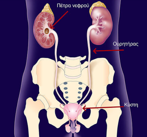
The kidneys are two bean-shaped organs located under the ribs towards the middle of the back, one on each side of the spine. The kidneys eliminate excess water and waste products from the blood, producing urine. They can also maintain a constant balance of salts and other substances in the blood. The kidneys produce hormones that help building strong bones and produce red blood cells. The tubes, called ureters, carry urine from the kidneys to the bladder, an oval-shaped chamber in the lower abdomen. Like a balloon, the elastic walls of the bladder stretch and extend to store urine. These walls contract when the bladder empties urine from the urethra outwards from the body.
What is kidney stone?
A kidney stone is a hard mass that grew from crystals separated from urine within the urinary tract. Normally, urine contains chemicals that prevent or inhibit crystal formation. These inhibitors don't seem to work well in everyone, so some people form stones. If the crystals remain small enough, they will travel through the urinary tract and pass out of the body in urine without being noticed.
Kidney stones can contain various combinations of chemicals. The most common type of stone is one that contains calcium in combination with either oxalates or phosphates. These chemicals are part of a person's normal diet and are found in important parts of the body, such as bones and muscles.
A less common type of kidney stone is caused by urinary tract infection. This type of stone is called a struvite stone or an inflammatory stone (from urinary tract infection). Another type of kidney stone is uric acid stones, which are slightly less common, while cystine stones are rare.
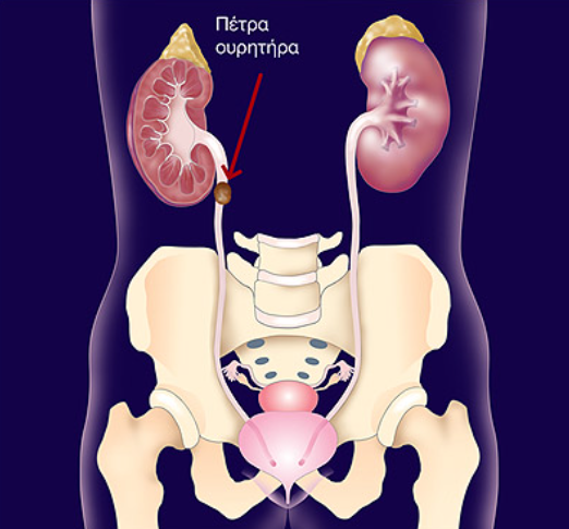
Nephrolithiasis is the medical term used to describe stones that form in the urinary tract. Doctors also use terms that describe the location of the stone in the urinary tract. For example, a stone in the ureter or ureterolithiasis is a kidney stone that descended into the ureter.
Gallstones and kidney stones are not related. They are in different areas of the body. Some with gallstones (gallstones) will not necessarily develop kidney stones.
Who has a kidney stone?
Stones occur more often in men. The prevalence of kidney stones increases dramatically as men enter their 40s and continues to rise into their 70s. For women, the prevalence of kidney stones peaks in their 50s.
Causes of kidney stones
Doctors do not always know what causes the formation of a stone. Although certain foods can help form stones in people who are sensitive, scientists do not believe that eating any food causes stones in people who are not sensitive.
A person with a family history of kidney stones may be more likely to develop stones. Urinary tract infections, kidney disorders, such as cystic degeneration of the kidneys, as well as certain metabolic disorders, such as hyperparathyroidism are associated with stone formation.
In addition, more than 70 percent of people suffering from a rare inherited disease called renal tubular acidosis develop kidney stones.
Cystinuria and hyperoxaluria are two other rare, inherited metabolic disorders that often cause kidney stones. In cystinuria, large amounts of the amino acid cystine, acid, which does not dissolve in the urine, is concentrated, leading to the formation of cystine stones. In patients with hyperoxaluria, the body produces too much oxalate. When urine contains more oxalate than can be dissolved, then these crystals precipitate in the form of stone.
Hypercalciuria is an inherited condition, which can be the cause of stones in more than half of patients. In absorbent hypercalciuria, excess calcium is absorbed by the body in excessive amounts from foods containing it and lost in urine. Another condition that leads to a high level of calcium in the urine is resorptive hypercalciuria where the kidney loses calcium in the urine. These high levels of calcium result in a high concentration in the urine of crystals of calcium oxalate or calcium phosphate capable of causing the formation of kidney stones or elsewhere in the urinary tract.
Similarly, hyperuricosuria — excess uric acid in the urine — which is a disorder of uric acid metabolism linked to gout or excessive consumption of protein products, can also cause kidney stones.
Other causes of kidney stones are excessive intake of vitamin D, urinary tract infections and urinary tract obstruction. Some pills such as diuretics and calcium-based antacids may increase the risk of kidney stone formation by increasing the amount of calcium in the urine.
Calcium oxalate stones may also occur in people who suffer from chronic bowel inflammation or who have undergone intestinal bypass surgery, or enterostomy surgery. As mentioned earlier, struvite stones can form in people who have had urinary tract infections. People taking the protease inhibitor, indinavir, a drug used to treat HIV infection, are at increased risk of developing kidney stones.
Which are the symptoms of kidney stones?
Kidney stones often cause no symptoms. Usually, the first symptom of a kidney stone is acute pain, which has been described as worse than labor pains, and which begins suddenly when a stone moves in the urinary tract and blocks the flow of urine. Usually, a person feels a sharp pain, like cramping in the back and ribs, in the area of the kidney or lower abdomen; Sometimes nausea and vomiting coexist. Later, the pain can spread to the groin.
If the stone is too large to pass easily, the pain continues as the muscles in the wall of the narrow ureter try to push the stone into the bladder. The combination of the movement of the stone and that the body tries to push it out, increases the likelihood that blood will appear in the urine, coloring the urine pink. As the stone moves down into the ureter and approaches the bladder, one may feel the need to urinate more often or feel a burning sensation when urinating.
If fever and chills accompany any of the above symptoms, then infection may coexist. In this case, the patient should immediately contact a doctor.
How is kidney stone diagnosed?
Sometimes the so-called "silent" kidney stone that does not cause symptoms, is discovered after random checking on X-rays taken during a general examination. If the stone is small, then it usually passes through the body unnoticed. Often, kidney stones are discovered after an X-ray or ultrasound done on someone visiting the emergency room for blood in the urine or sudden pain. These diagnostic tests will give the doctor valuable information about the size and location of the stone. Blood and urine tests may help identify any abnormal substances that could facilitate kidney stones.
The doctor may decide to scan the urinary tract using a special test called computed tomography (CT) or intravenous pyelography (IVP) and lately the combination of the two above tests called CT pyelography. The results of all these tests will help determine the appropriate treatment.
Prevention of kidney stones
A person who has had a kidney stone more than once is likely to have it in the future. So, if possible, prevention is important. To determine the cause of the formation of the kidney stone, the doctor will order certain laboratory tests, including urinalysis and blood tests. The doctor will ask questions about the medical history, the profession of the patient, as well as his eating habits; If a stone has been removed endoscopically, or if the patient has expelled a stone and was able to collect it, then laboratory analysis of the stone can help the doctor plan treatment.
The doctor may ask the patient to collect urine for 24 hours after a stone has passed on its own or has been removed endoscopically. For a 24-hour urine collection, the patient urinates in a large container, which is kept in the refrigerator between urinations. The collection is used to measure urine volume and acidity levels, as well as calcium, sodium, uric acid, oxalic, citric acid and creatinine, a product of muscle metabolism. The doctor will use this information to determine the cause of the formation of the kidney stone. A second 24-hour urine collection may be needed to determine if the treatment is working.
How are kidney stones treated?
Usually, surgery is not necessary. Most kidney stones can pass through the urinary tract with plenty of water — 2 to 3 liters a day — with the aim of causing frequent urination and eliminating the stone along with urine. Often, the patient has to stay at home during this procedure, drinking fluids and taking painkillers, as needed. The doctor usually asks the patient to collect the stone when it passes along with the urine.
Lifestyle changes
A simple and most important lifestyle change to prevent kidney stone formation is to drink more fluids (water is best). Someone who tends to have stones should try to drink enough fluids throughout the day to produce at least 2–3 liters of urine every 24 hours.
In the past, people who had calcium stones advised them to avoid dairy products and other foods high in calcium. Recent studies have shown that calcium-rich foods, such as dairy products, may help prevent calcium stone formation. However, taking calcium in pill form can increase the risk of developing stones.
Patients should avoid foods with added vitamin D and certain types of calcium-based antacids. Someone who has highly acidic urine may need to eat less meat, fish, and poultry. These foods increase the amount of acid in the urine.
To prevent cystine stones, a person should drink enough water each day to dissolve the concentration of cystine, which is excreted in the urine, which can be difficult. More than 3 litres of water may be required 24 hours a day, and a third of this should be drunk overnight.
Drug Therapy
A doctor may prescribe some medications to help prevent the formation of calcium stones and uric acid. These drugs control the amount of acid or alkali in the urine, key factors in crystal formation. Allopurinol is a drug that may also be useful in some cases of hyperuricosuria.
Doctors usually try to control hypercalciuria — to prevent the development of calcium stones — by prescribing certain diuretics, such as hydrochlorothiazide. These drugs reduce the amount of calcium released by the kidneys in the urine, favoring the retention of calcium in the bones. They work best when sodium (salt) intake is low.
Rarely, patients with hypercalciuria can be given cellulose with sodium phosphate, which binds calcium in the intestine and thus prevents its leakage into the urine.
For struvite stones that have been completely removed, the first line of prevention is to keep urine free of bacteria that can cause infection. The patient's urine should be checked regularly to ensure that bacteria are not present.
Patients with hyperparathyroidism sometimes develop calcium stones. Treatment in these cases is usually surgical removal of the parathyroid glands, located in the neck. In most cases, only one of the glands swells. Removal of glands treats the patient's problem with hyperparathyroidism and kidney stones.
Surgical treatment
Surgery may need to remove a kidney stone, in cases of:
- if it is not eliminated on its own after a reasonable period of time and causes constant colic
- if it is too big to pass on its own
- if it is found in a difficult position
- if it obstructs urine flow
- if it causes continuous urinary tract infections
- if it causes damage to kidney tissue or continuous bleeding
- if its volume has increased, on control X-rays
Until 20 years ago, open surgery was necessary to remove a stone. Surgery requires a recovery time of 4 to 6 weeks. Today, this remedy is used only in very rare cases of complications.
Extracorporeal shockwave lithotripsy
Extracorporeal shockwave lithotripsy (ESWL) is the most commonly used procedure for the treatment of kidney stones. In ESWL, shock waves generated outside the body travel through skin and body tissues until they reach the densest stones. The stones break down into small particles that make it easier for them to pass through the urinary tract with urine.
Several types of ESWL devices exist. Most devices use either X-rays or ultrasound to help the surgeon locate the stone during the procedure. For most types of extracorporeal lithotripsy procedures, anesthesia is not required.
In most cases, extracorporeal lithotripsy can be performed on an outpatient basis. Recovery time is relatively short, and most people can return to normal activities within a few days.
Complications can occur with ESWL. Some patients have blood in their urine for a few days after treatment. Bruising and minor discomfort in the back or abdomen may be observed from shock waves. To reduce the risk of complications, doctors typically tell patients to avoid taking aspirin and other medications that affect blood clotting for several weeks before treatment.
Sometimes, broken stone fragments cause minor blockage as they pass through the urinary tract and can cause discomfort. In some cases, the doctor will insert a small tube called a stent or pig tail through the bladder into the ureter to help the fragments descend. Sometimes the stone is not completely crushed with one session, and additional sessions may be needed.
As with any invasive surgical procedure, you should discuss with your doctor the potential risks and complications before making a treatment decision.
In this procedure, the urologist surgeon makes a small incision of 1 cm in the back and creates a tunnel into the kidney. Using a tool called a nephroscope, surgeons locate and remove or crush the stone into small pieces with the help of laser. Often, patients stay in the hospital for a few days and may have a small tube called a nephrostomy tube in the kidney during the procedure healing.
One advantage of percutaneous nephrolithotripsy is that the surgeon can remove some of the stone pieces immediately, rather than relying solely on their natural passage through the kidney.
Removal of the stone endoscopically
Although some ureter stones may be treated with extracorporeal lithotripsy (ESWL), ureteroscopy may be required to remove a stone through the ureter. No incision is made in this procedure. Instead, the surgeon passes a small fiber-optic instrument (which has a small video camera) called a ureteroscope through the urethra and bladder into the ureter.
The surgeon then places the stone and removes it with forceps or crushes it with the help of laser. A small pig tail or stent can be left in the ureter for a few days to help urine flow.
Hope through research
New drugs and the growing field of lithotripsy have significantly improved the treatment of kidney stones. Researchers are also trying to answer questions such as:
- Why do some people continue to have stones that cause pain (colic)?
- How is it possible for medicine to predict, which patients have an increased risk to form stones?
- What are the long-term effects of lithotripsy?
- What genes play a role in stone formation?
- What is the natural substance(s) found in urine that inhibit stone formation?
Researchers are also developing new drugs with fewer side effects.
Points to keep in mind
- A person with a family history of lithiasis or a personal history of more than one episode of kidney stones is more likely to develop stones again.
- A good first step that can prevent the formation of any kind of stone is to drink plenty of fluids — water is best.
- Someone who has an increased risk for developing stones may need certain blood and urine tests to determine what factors can be changed to reduce this risk.
- Some individuals will need medications to prevent stone formation.
- People with chronic urinary tract infections have an increased risk of developing kidney stones and will often need to have the stone removed if the doctor determines that the stone is causing the infection. Patients should have regular postoperative monitoring to be sure that the infection has been eradicated.
Kidney Lithiasis (kidney stone)
Kidney stones (kidney stones, nephrolithiasis) are hard deposits of minerals and salts that form inside your kidneys. The causes are many and can affect any part of your urinary tract (from your kidneys to the bladder). Often, stones form when urine stagnates, allowing mineral salts to crystallize and stick together.
The passage of kidney stones can be quite painful, but stones usually do not cause permanent damage if they are recognized in time. Depending on your condition, you may need more than just taking pain medication and drinking lots of water to eliminate a kidney stone. If stones remain in the urinary tract, they can cause serious urinary tract infections of the kidneys (eg, pyelonephritis) or other complications — surgery may be needed.
Your doctor may recommend preventive treatment to reduce the risk of recurrent kidney lithiasis if there is an increased risk of it occurring again.
Symptoms
A kidney stone may not cause symptoms until it moves into your kidney or passes into your ureter — the tube that connects your kidney and bladder.
- Severe pain in the side and back, under the ribs
- Radiating pain in the lower abdomen and groin
- Pain that comes in waves and increases in intensity
- Pain when urinating
- Pink, red or brown urine
- Cloudy urine or odor
- Nausea and vomiting
- Persistent urge to urinate
- Urination more frequent than usual and little amount
- Fever and chills if there is an infection
The pain caused by a kidney stone can change — for example, changing position or increasing tension — as the stone moves through your urinary tract.
When to see a doctor
Make an appointment with your doctor if you have symptoms that worry you.
Seek immediate medical attention if you experience:
- Pain so severe that you cannot sit or find a position that relieves you
- The pain is accompanied by nausea and vomiting
- The pain is accompanied by fever and chills
- Blood in urine
- Difficulty urinating
Causes
Kidney stones often have no specific cause, although many factors may increase the risk.
Kidney stones form when your urine contains more crystal-forming substances — such as calcium, oxalic and uric acid. At the same time, your urine may lack substances that prevent crystals from sticking together, creating an ideal environment for kidney stones to form.
Types of kidney stones
Knowing the type of kidney stone helps determine the cause and can give clues on how to reduce the risk of more kidney stones forming. If possible, try to collect the stone if you miscarry one so you can bring it to your doctor for analysis.
Types of kidney stones include:
- Most kidney stones are calcium stones, usually in the form of calcium oxalate. Oxalate is a naturally occurring substance found in food and is also created daily by your liver. Some fruits and vegetables, as well as nuts and chocolate, are high in oxalates.
- Dietary factors, high doses of vitamin D, intestinal bypass surgery, and several metabolic disorders can increase the concentration of calcium or oxalate in the urine.
- Stone stones. Struvite stones form as a result of an infection, such as a urinary tract infection. These stones can grow quickly and become quite large, sometimes with few symptoms or little warning.
- Uric acid stones. Uric acid stones can form in people who don't drink enough fluids or lose a lot of fluids, those who eat high-protein foods and those who have gout.
- Cystine stones. These stones form in people with an inherited disorder that causes the kidneys to secrete too many amino acids (cystinuria).
Risk factors
Factors that increase the risk of developing kidney stones include:
Family or personal history
If someone in your family has kidney stones, you're more likely to develop them too, and if you already have one or more kidney stones, there's an increased risk of developing another.
Dehydration
Not drinking enough water each day can increase your risk for kidney stones. People who live in warm climates and those who sweat a lot may be at greater risk than others.
Certain diets
Eating a diet high in protein, sodium (salt) and sugar can increase the risk of certain forms of kidney stones. Too much salt in your diet increases the amount of calcium your kidneys need to filter through and significantly increases your risk for kidney stones.
Obesity
High body mass index (BMI), large waist circumference and weight gain have been linked to an increased risk of kidney stones.
Digestive diseases and surgery
Gastric bypass surgery, inflammatory bowel diseases or chronic diarrhea can cause changes in the process that affect calcium and water absorption, increasing levels of these substances and forming stones in your urinary tract.
Other medical conditions
Diseases and conditions that may increase your risk for kidney stones include renal tubular acidosis, cystinuria, hyperparathyroidism, certain medications, and certain urinary tract infections.
Contents
Book an Appointment
Dr. Mertziotis specialises in diagnosing and treating kidney stones using modern methods including ESWL, percutaneous nephrolithotripsy, and endoscopic techniques.
Book an Appointment Online +30 210 6465359